Title
The Effect of Polyethylene Glycol on Polycaprolactone Electrospun Scaffolds: Morphology, Mechanical Properties, and Nerve Growth Factor Delivery Profile
Authors
Manuel A. Alfaro De Pra,a,b Daniela S. Coelho,a Rosa M. Ribeiro-do-Valle,a J. A. Lopes-da-Silva,c M. Maraschin*a and Beatriz Veleirinho*ab
aNanoBioMat, Federal University of Santa Catarina, 88040-900, Florianópolis, Brazil.
bNanoScoping- Solutions in Nanotechnology, 88040-400 Florianópolis, Brazil.
cQOPNA Research Unit, Department of Chemistry, University of Aveiro, 3810-193, Aveiro, Portugal.
*Corresponding author E-mail address: bveleirinho@gmail.com (B. Veleirinho), m.maraschin@ufsc.br (M. Maraschin)
Article History
Publication details: Received: 17th June 2020; Revised: 16th July 2020; Accepted: 16th July 2020; Published: 04th August 2020
Cite this article
de Pra M.A.A.; Coelho D.S.; Ribeiro-do-Valle R.M.; Lopes-da-Silva J. A.; Maraschin M.; Veleirinho B. The Effect of Polyethylene Glycol on Polycaprolactone Electrospun Scaffolds: Morphology, Mechanical Properties, and Nerve Growth Factor Delivery Profile. Nano Prog., 2020, 2(4), 24-31.
Abstract
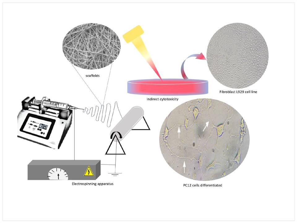
Electrospinning is a promising technique to generate scaffolds for delivery systems and nerve tissue engineering. This study aimed to investigate the suitability of polyethylene glycol (PEG) as an agent to prolong circulation time in the delivery of nerve growth factor (NGF) encapsulated into polycaprolactone (PCL) electrospun fibers. Three molecular weights (400, 1500, and 35000 Da) and concentrations (1, 5, and 10% ratio to PCL) of PEG were studied. The effect on NGF from the scaffolds was evaluated by neuronal differentiation using PC12 cells. Scaffolds were characterized for their architecture by Scanning Electron Microscopy (SEM) and for their mechanical properties. Cytotoxicity was evaluated by MTT and agarose overlay methods. The effect of fiber architecture on PC12 morphology was also investigated. Nanofibrous hybrid mats were obtained for all PCL/PEG blends tested. The addition of PEG caused a reduction in the average due to the increased stretching ability over the electrospinning process. This reduction on fiber diameter showed impact on mechanical properties of the fibrous scaffolds, i.e. lower tensile strength and elongation were observed for scaffolds with 5% and 10% PEG. The co-lyophilization of NGF with PEG prior to nanofiber production improved the delivery profile of NGF, stimulating neuronal cell differentiation even after 28 days of continuous release.
Keywords
Electrospinning; scaffold; polycaprolactone; growth factor delivery; nerve repair


